Promotional Features


Lighting up a new pathway for positive skin aging - through autophagy and proteasome activity
Globally, by the year 2030, 1 in 6 people will be over 60 years of age. Estimates, from the World Health Organization, indicate that within the next 30 years, this portion of the population will double from 1 billion people in 2020 to 2.1 billion in 20501.
Aging is the next frontier in the debate surrounding inclusion and diversity. Stereotypes persist in today's society, and younger people view aging with anxiety and fear. However, a new generation of seniors is starting to reject this view through a more positive approach.
Studies have shown that older people who view their age in a positive light recover faster from illness or mobility problems and are less likely to suffer from dementia.
The increase in life expectancy and the impact of aging on the biological function and appearance of the skin is directly proportional to the increased demand for dermatologists, beauticians, plastic surgeons and pro-aging products. Individuals are motivated by their natural desire to live longer as well as to remain, and appear, healthy2,3.
The face and hands are the regions of the body where signs of aging first appear4,5. These changes are manifested by the increase of skin wrinkles, sagging, and dryness, as well as the appearance of age spots6,7.
Age spots, also known as solar lentigines and senile lentigines, are light brown to black pigmented lesions of different sizes that become more evident in individuals 40-50 years old and gradually increase with age in regions such as the face, back, hands, arms and shoulders.
The best explanation for the appearance of spots on the skin are the formation and accumulation of melanin and lipofuscin pigments. However, the increase or decrease of melanin in the skin tissue does not occur as result of aging, while increasing age is a determining factor for the appearance of lipofuscin in the epidermis and dermis8. Thus, it is possible to say that age spots are not exactly the result of melanin accumulation, but likely include the presence of lipofuscin – excess of oxidized or damaged proteins, lipids and other cellular residues in lysosomes, cellular organelle responsible for organic substances digestion9.
Lipofuscin, from Greek "lipo" which means fat and from Latin "fuscus" which means dark, is a yellowish-brown pigment found in various cells of the body, such as keratinocytes, fibroblasts, neurons, among others. Its abundance and availability are greater in elderly individuals than in young people, which connotes the popular term “aging pigment” to this component and allows its adoption as a biomarker for the determination of absolute age10.
Although senile spots are more evident in individuals over 40-50 years old, recent studies have shown that lipofuscin – or cellular waste – accumulates inside the cell in individuals from 30-35 years of age, a stage in which the presence of residue exceeds the cell's ability to catabolize and recycle this waste11.
Many physiological functions are dependent on the degradation and recycling of damaged molecules within the cell12 and the processes that promote the supply of components necessary for the replacement of cellular energy are autophagy and proteasome activity. The autophagic process and proteasome activity are distinct but complementary phenomena. Both of these metabolic pathways specialize in degrading their respective targets. While autophagy acts by recycling macromolecules such as old organelles and damaged macroproteins, proteasome activity has the function of digesting small damaged proteins13.
Iselight is a new product launch from Chemyunion that stimulates these two cellular self-cleaning processes, while preventing the formation of more residue inside the cell, and rescuing cellular homeostasis. In this way, it acts to reduce age spots in addition to improving the texture and elasticity of the skin.
Iselight is a synergistic combination of Myrothamnus flabellifolia extract, a resurrection plant known in traditional African medicine, and upcycled Coffea arabica extract, an important source of antioxidants, which was obtained after numerous ingredient screenings and screening tests to achieve the precise composition required to best stimulate autophagy and proteasome activity.
Lipofuscin Reduction
To assess the ability of Iselight to decrease Lipofuscin, human fibroblasts were treated and incubated for 24 hours for subsequent irradiation with three doses of UVA per day, totaling 7J/cm2. After this period, the cells were submitted to the in vitro immunofluorescence technique by a lipofuscin detection method using antibody.
The irradiated and active-treated group was compared to the control group (no treatment) and the irradiated control group (no treatment and irradiated with UVA 7J/cm2).
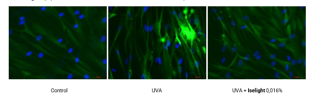
Figure 1: Immunofluorescence analysis of Lipofuscin (green) and DAPI counterstaining (blue, cell nuclei marker), microscopic images at 40x magnification.
As can be seen in the Figure 1, Iselight a was able to eliminate lipofuscin generated by exposure to UVA radiation, matching the control group without irradiation stress.
Clinical trial
The clinical study was conducted with a panel of 63 female volunteers between the ages of 45 and 69 years (mean age 57 years), presenting with photoaging spots on the hands and facial photoaging (facial sagging, wrinkles/fine lines and blemishes). The skin tone lightening activity of 2% Iselight was evaluated compared to placebo, under normal conditions of use, applied twice a day on back of hands and face. Analysis times were day 0 (before treatment), after 28 days and after 56 days of treatment.
Evenness skin tone on the hands
The analysis of spots on the back of hands was performed using standardized images captured with a photographic camera and instrumental colorimetry measurements with the Chroma Meter CR 400 Konica Minolta equipment. All analyses were accompanied by a dermatologist.
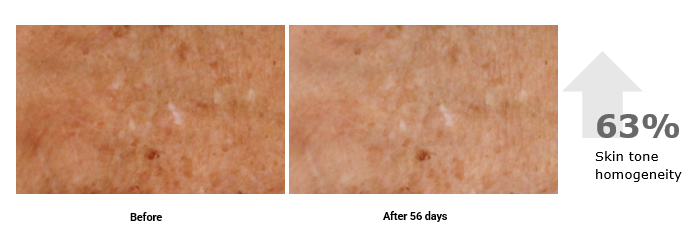
Figure 2: Lightening and evenness skin tone on hands before and after 56 days of treatment with Iselight 2%.
The results obtained through standardized images showed an average increase of 44.7% and 63.2% in the skin tone homogeneity on hands after, respectively, 28 and 56 days of treatment with Iselight.
-----
References:
- World Health Organization
- YAAR, Mina. Clinical and histological features of intrinsic versus extrinsic skin aging. In: Skin aging. Springer, Berlin, Heidelberg, 2006. p. 9-21.
- MAKRANTONAKI, E; Zouboulis, C. Characteristics and pathomechanisms of endogenously aged skin. Dermatology, v. 214, n. 4, p. 352-360, 2007.
- GOLD, Michael H.; GALLAGHER, Conor. An evaluation of the benefits of a topical treatment in the improvement of photodamaged hands with age spots, freckles, and/or discolorations. Journal of drugs in dermatology: JDD, v. 12, n. 12, p. 1468-1472, 2013.
- FARAGE, Miranda A. et al. Structural characteristics of the aging skin: a review. Cutaneous and ocular toxicology, v. 26, n. 4, p. 343-357, 2007.
- RINNERTHALER, Mark et al. Oxidative stress in aging human skin. Biomolecules, v. 5, n. 2, p. 545-589, 2015.
- YAMAGUCHI, Yuji; BRENNER, Michaela; HEARING, Vincent J. The regulation of skin pigmentation. Journal of biological chemistry, v. 282, n. 38, p. 27557-27561, 2007.
- PORTA, Eduardo A. Pigments in aging: an overview. Annals of the New York Academy of Sciences, v. 959, n. 1, p. 57-65, 2002.
- SKOCZYŃSKA, Anna et al. Melanin and lipofuscin as hallmarks of skin aging. Advances in Dermatology and Allergology/Postȩpy Dermatologii i Alergologii, v. 34, n. 2, p. 97, 2017.
- BOURNE, Geoffrey H. Lipofuscin. In: Progress in brain research. Elsevier, 1973. p. 187-201.
- RÜBE, Claudia E. et al. Human skin aging is associated with increased expression of the histone variant H2A. J in the epidermis. npj Aging and Mechanisms of Disease, v. 7, n. 1, p. 1-11, 2021.
- HEWITT, Graeme; KOROLCHUK, Viktor I. Repair, reuse, recycle: the expanding role of autophagy in genome maintenance. Trends in cell biology, v. 27, n. 5, p. 340-351, 2017.
- NAM, Taewook et al. Emerging paradigm of crosstalk between autophagy and the ubiquitin-proteasome system. Molecules and cells, v. 40, n. 12, p. 897, 2017.